Orthopedic Insights & Knowledge
Explore the latest advances in orthopedic implants, trauma solutions, surgical technology, and healthcare industry news from Auxein Medical.
Choosing a Pedicle Screw System in 2026 requires more than comparing product brochures. The right decision begins with the patient’s anatomy, surgical objective, and risk profile. A screw designed for a short lumbar construct may not suit a long thoracolumbar fusion. A smooth operating-room workflow can matter as much as a clever feature.
Dr. Christopher I. Shaffrey, a widely recognized spine surgeon and researcher, has emphasized, “The goal is to get patients back to a normal life.” That principle keeps hardware selection grounded. Surgeons should examine screw diameter, length options, thread design, tulip strength, rod compatibility, reduction capability, and implant materials. Navigation and robotic integration may improve placement confidence, but technology cannot replace anatomical judgment. Not every hospital has the same imaging access, training resources, or technical support.
In 2026, evaluation should also include clinical evidence, manufacturing consistency, sterilization processes, regulatory status, and long-term revision considerations. A system may perform well in a controlled study yet create difficulties during a crowded revision procedure. That detail is easy to overlook. Hands-on trial experience, peer feedback, and transparent supplier support can reveal practical weaknesses before they affect patients.
No checklist is perfect. Surgeons can still choose poorly when marketing language overwhelms evidence. This guide examines the questions that deserve closer attention before selecting a Pedicle Screw System. It considers safety, efficiency, compatibility, cost, and adaptability without pretending that one system fits every spine.
What Is a Pedicle Screw System and How Does It Work?
How to Choose a Pedicle Screw System in 2026?
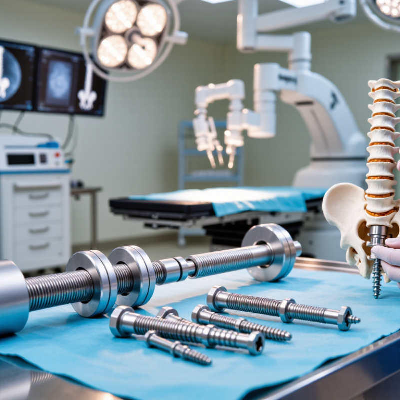
A pedicle screw system stabilizes the spine during selected surgical procedures. It usually includes screws, rods, connectors, and locking components. The surgeon places each screw through the vertebral pedicle into the stronger central bone. Rods then link the screws and help control movement between spinal segments. This structure can support alignment while bone fusion develops. It is not a replacement for careful diagnosis.
The working principle is mechanical. Properly positioned screws resist bending, rotation, and shifting forces. Screw diameter, length, thread design, and trajectory must match the patient’s anatomy. Imaging helps assess bone quality, deformity, and nearby nerves. In practice, navigation may improve accuracy, but it cannot replace surgical judgment. No system is perfect. A neat-looking construct may still be unsuitable.
Tips: Check compatibility between screws, rods, and instruments. Review imaging before selecting sizes. Consider osteoporosis, revision surgery, and unusual anatomy. Ask whether the system offers reliable locking and clear visual feedback. Small details matter. The final choice should follow current clinical evidence, regulatory requirements, and the surgeon’s trained assessment. A second review can expose assumptions that the first plan missed.
How to Choose a Pedicle Screw System in 2026? - What Is a Pedicle Screw System and How Does It Work?
| Evaluation Dimension | What It Is / How It Works | Common Options | Selection Considerations | Verification Points |
|---|---|---|---|---|
| Primary Function | Anchors are inserted through the vertebral pedicles and connected with rods to stabilize, align, or correct a spinal segment. | Posterior fixation, deformity correction, fusion support, and fracture stabilization. | Match the system to the spinal region, pathology, surgical objective, and planned construct length. | Confirm the intended indication, anatomical coverage, and available implants before surgery. |
| Screw Diameter and Length | The screw transfers load from the rod to the vertebra through purchase in the pedicle and vertebral body. | Multiple diameters and lengths designed for thoracic, lumbar, and sacral anatomy. | Select dimensions that fit the pedicle and provide adequate bone purchase while avoiding cortical breach or neural and vascular structures. | Review CT or other appropriate imaging, anatomical measurements, and the system’s size range. |
| Screw Design | Thread geometry and tip design help the screw engage cancellous and cortical bone. | Standard, reduction, cannulated, fenestrated, fixed-angle, and polyaxial screws. | Polyaxial heads can simplify rod alignment; reduction screws may assist controlled translation; fenestrated designs may be used with bone cement in selected cases. | Check approved indications, compatibility with instruments, and cement-use instructions where applicable. |
| Rod Options | Rods link the screw heads and help maintain alignment while the fusion or healing process occurs. | Straight or pre-contoured rods; commonly available in titanium alloy or cobalt-chromium alloy. | Consider contouring needs, stiffness, correction goals, rod diameter, and compatibility with screw heads and connectors. | Confirm that rods, set screws, cross-links, and connectors belong to the same validated system. |
| Material Selection | Implant materials provide structural strength and corrosion resistance for long-term internal fixation. | Titanium alloy, cobalt-chromium alloy, and selected radiolucent or polymer components for specific applications. | Evaluate strength, stiffness, imaging characteristics, metal sensitivity history, and compatibility with other implants. | Review material specifications, corrosion data, MRI conditions, and applicable regulatory documentation. |
| Open or Percutaneous Access | Open systems are placed through direct exposure, while percutaneous systems use guidewires, extensions, and smaller access channels. | Open instrumentation, minimally invasive instrumentation, or a modular system supporting both techniques. | Choose according to surgical approach, imaging workflow, soft-tissue objectives, training, and available operating-room equipment. | Assess guidewire control, screw extension stability, targeting accuracy, and instrument ergonomics. |
| Deformity Correction Features | Correction is achieved through controlled rod placement, screw reduction, compression, distraction, and rotational maneuvers. | Reduction towers, derotation tools, multi-rod connectors, domino connectors, and transverse connectors. | For complex deformity, prioritize a broad implant range, predictable reduction control, and stable locking mechanisms. | Review published mechanical testing, correction technique instructions, and compatibility of correction instruments. |
| Bone Quality Adaptability | Fixation depends on the interaction between screw threads and available bone, which can be reduced in osteoporosis or revision cases. | Cement-augmented options, larger-diameter screws, expandable designs, and supplemental fixation strategies. | Use only when clinically appropriate and follow augmentation limits, cement-handling requirements, and imaging guidance. | Evaluate bone density, purchase quality, cement leakage risk, and the system’s specific contraindications. |
| Imaging and MRI Conditions | Implant material and geometry can affect artifact levels and determine the conditions under which MRI may be performed. | MRI-conditional systems with specified field strength, spatial-gradient, SAR, and scan-duration limits. | Select a system whose imaging profile supports postoperative assessment and whose MRI conditions are practical for the patient population. | Read the official MRI safety information; do not assume that all metallic implants have identical conditions. |
| Instrument Workflow | Drivers, taps, awls, guides, towers, and rod inserters determine how efficiently and consistently the system can be used. | Manual, navigated, robotic-compatible, open, minimally invasive, and modular instrument sets. | Prioritize intuitive assembly, tactile feedback, clear size markings, low instrument complexity, and reliable sterilization processes. | Check trial availability, instrument completeness, cleaning instructions, and staff training requirements. |
| Mechanical and Clinical Evidence | Mechanical tests assess properties such as static strength, fatigue strength, and connection security; clinical evidence evaluates outcomes and complications. | Bench testing, cadaveric evaluation, prospective studies, retrospective studies, and post-market surveillance. | Compare evidence quality, follow-up duration, patient population, complication reporting, and relevance to the intended indication. | Request published data and regulatory documentation rather than relying only on marketing claims. |
| Sterilization and Reprocessing | Reusable instruments must be cleaned, inspected, packaged, and sterilized according to validated instructions. | Reusable instrument trays, limited-use components, and sterile single-use accessories. | Consider turnaround time, tray weight, maintenance requirements, inspection access, and hospital sterilization capacity. | Verify validated cleaning and sterilization parameters and inspect instruments for wear or damage. |
| Regulatory and Supply Support | Availability of approved implants, instruments, technical documentation, training, and post-market support affects implementation reliability. | Regulatory clearance or approval, traceability, inventory support, technical training, and case coverage. | Choose a system with dependable supply, documented quality controls, responsive technical support, and transparent complication reporting. | Confirm current authorization, lot traceability, shelf-life information, availability of replacement components, and local service capability. |
Note: Final implant selection should be made by a qualified spine surgeon based on patient anatomy, diagnosis, bone quality, imaging, surgical approach, and the current instructions for use.
FAQS
It is a spinal fixation system used during selected surgical procedures. It usually includes screws, rods, connectors, and locking parts.
Screws pass through vertebral pedicles into stronger central bone. Rods connect the screws and limit unwanted movement between spinal segments.
It may support unstable segments caused by deformity, traumatic fracture, selected tumors, infection, or failed previous fusion.
No. Back pain alone is not enough. Imaging, symptoms, examination findings, and functional limits must support the decision.
Diameter and length should match pedicle width, bone quality, trajectory, and surgical goals. A larger screw is not automatically safer.
Compare titanium and cobalt-chrome options, head movement, thread shape, shaft stiffness, and insertion feel. No design is universally better.
No. These tools may improve placement accuracy, but trained surgical judgment remains essential.
Confirm compatibility among screws, rods, connectors, and instruments. Review imaging, bone density, unusual anatomy, and regulatory information.
A neat construct may still be unsuitable. Healing can vary, and unfamiliar instruments may slow placement. A second review can reveal missed assumptions.
Conclusion
Choosing the right Pedicle Screw System in 2026 requires a balanced evaluation of clinical purpose, design, compatibility, and long-term value. These systems stabilize the spine by anchoring screws into the vertebral pedicles and connecting them with rods, helping restore alignment and support fusion. They may be considered for conditions such as spinal instability, deformity, fractures, degenerative disease, or certain revision procedures, depending on the patient’s diagnosis and treatment plan.
When comparing options, assess screw design, materials, diameter, length, thread pattern, fixation strength, and available adjustment features. Surgical compatibility is equally important, including instrument integration, imaging requirements, navigation support, and ease of placement. Patient-specific factors such as bone quality, anatomy, age, activity level, and medical history should guide selection. Finally, review safety data, manufacturing quality, regulatory compliance, total procedure cost, durability, imaging considerations, and follow-up performance. The final choice should be made by qualified spinal professionals through careful clinical assessment and shared decision-making.