Orthopedic Insights & Knowledge
Explore the latest advances in orthopedic implants, trauma solutions, surgical technology, and healthcare industry news from Auxein Medical.
How to Choose the Right TLIF Lumbar Cage?
Table of Contents
- Understanding TLIF Lumbar Cage Design and Surgical Purpose
- Assessing Patient Anatomy, Diagnosis, and Spinal Stability
- Comparing Cage Materials, Shapes, Sizes, and Lordotic Angles
- Matching Cage Selection to Surgical Technique and Fusion Goals
- Reviewing Safety, Biocompatibility, and Long-Term Clinical Needs
- FAQS
- Conclusion
- Related Posts
Choosing the right TLIF Lumbar Cage is a clinical decision, not a simple product comparison. The cage must support spinal stability, encourage bone growth, and fit the patient’s anatomy. It should also match the surgeon’s technique, imaging findings, and long-term treatment plan.
A reliable choice begins with careful assessment. Surgeons may review disc height, foraminal narrowing, bone quality, spinal alignment, and previous surgery. Cage size, shape, material, lordotic angle, and insertion method can all affect placement. A cage that looks suitable on a catalog page may not suit a narrow disc space or weakened vertebral endplates. Small differences matter.
Not every patient needs the same design.
Common materials include PEEK, titanium, and composite structures. Each option has potential strengths and limitations involving imaging, surface integration, stiffness, and handling. Clinical evidence can guide the discussion, but it does not replace professional judgment. The surgeon’s experience with a specific TLIF Lumbar Cage also matters, especially during challenging minimally invasive procedures.
This guide explains the practical factors behind cage selection, including anatomy, biomechanics, graft compatibility, radiographic assessment, and manufacturer quality controls. It also considers risks such as subsidence, migration, nonunion, and imperfect positioning. These outcomes are not always predictable. A thoughtful decision recognizes uncertainty instead of promising guaranteed fusion.
Patients should discuss their diagnosis, bone health, surgical goals, and available cage options with a qualified spine specialist. The best choice is usually the one that fits the clinical situation, not the one with the longest feature list. Expect careful questions. Even experienced teams sometimes reassess their preferred design.
Understanding TLIF Lumbar Cage Design and Surgical Purpose
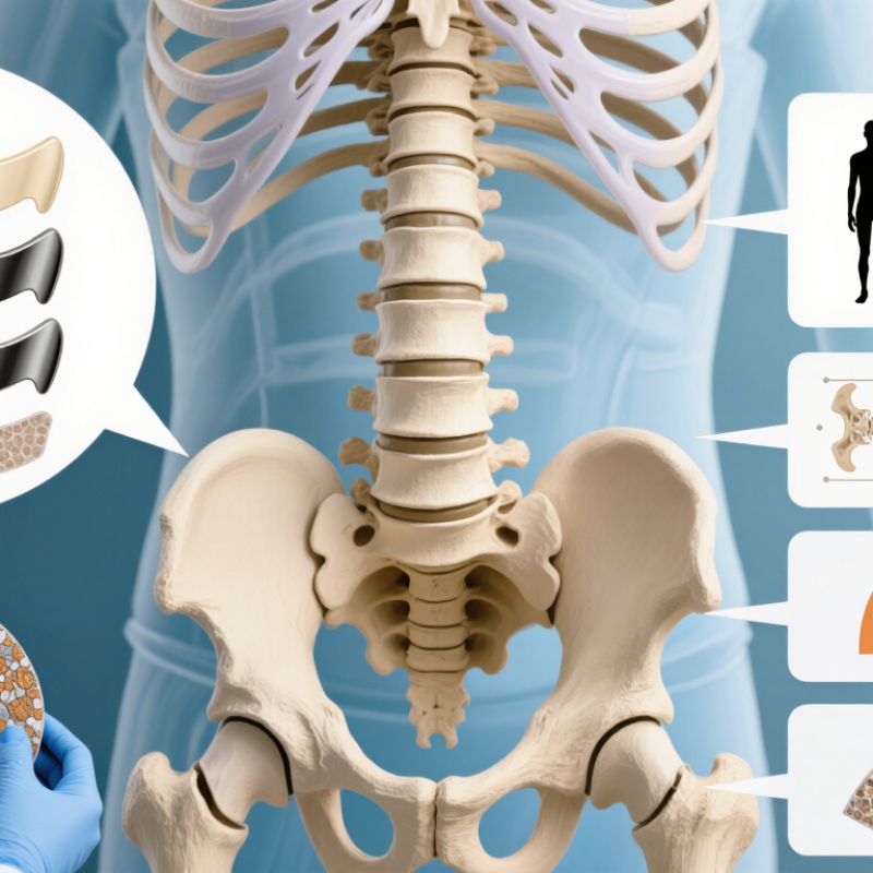
Choosing a TLIF lumbar cage begins with surgical purpose, not a catalogue image. The cage should restore disc and foraminal height, support anterior column loading, and help maintain segmental alignment after decompression. A wider footprint may reduce endplate stress, while excessive height can injure the endplate or create subsidence. Small details matter.
Cage geometry should match the patient’s anatomy and the surgeon’s insertion route. Consider footprint, lordotic angle, graft volume, radiographic visibility, and endplate preparation. Computed tomography can reveal bone quality and endplate shape; magnetic resonance imaging clarifies neural compression. The 2023 World Health Organization report identifies osteoporosis as a major global health burden, so bone strength deserves attention before implantation. Poor bone quality may require different height selection and fixation planning.
Clinical evidence is encouraging, but not perfect. A 2022 systematic review in Global Spine Journal reported generally high fusion rates after TLIF, often above 90%, although study definitions and follow-up periods differed. That variation matters.
A radiographic fusion does not always equal pain relief or restored function. In practice, cage selection should be discussed alongside bone density, sagittal alignment, symptoms, and the planned decompression. No single design fits every spine. Even experienced teams must reassess their assumptions.
Assessing Patient Anatomy, Diagnosis, and Spinal Stability
How to Choose the Right TLIF Lumbar Cage?
Patient anatomy should guide cage selection, not catalog dimensions. Measure disc height, foraminal width, endplate shape, and segmental lordosis on CT and MRI. A narrow cage may miss the strongest endplate zone. An oversized cage may cause distraction, subsidence, or nerve irritation. Endplate preservation matters. So does bone quality.
Diagnosis changes the surgical target. Foraminal stenosis may require height restoration, while stable degenerative disc disease may need less correction. In spondylolisthesis, assess slip severity, facet condition, and flexion-extension motion. A 2022 systematic review in Global Spine Journal reported TLIF fusion rates commonly near 90–95%, although cage designs and fusion definitions varied. That number is useful, but not absolute.
Spinal stability remains the difficult judgment. Consider instability, sagittal imbalance, osteoporosis, and adjacent-level stress. DEXA, CT attenuation, and clinical risk factors can reveal weak fixation conditions. The cage should support load sharing without forcing excessive lordosis. Short-term radiographs can look excellent. Later subsidence may tell another story. I would not pretend one cage height fits every patient. Surgical experience, careful trialing, and postoperative imaging still matter. Evidence guides the choice, but anatomy makes the final decision.
Comparing Cage Materials, Shapes, Sizes, and Lordotic Angles
How to Choose the Right TLIF Lumbar Cage?
Cage selection should match anatomy, bone quality, and the desired spinal profile. PEEK cages offer radiolucent imaging, making bone growth easier to assess on follow-up scans. Titanium cages provide strong surface integration, but they can create more imaging artifact. Hybrid designs may balance both advantages. A 2022 systematic review in The Spine Journal reported fusion rates commonly above 90% in modern TLIF studies, although definitions varied between trials.
Shape matters at the disc space. Bullet-shaped cages may ease insertion, while wider footprints can reduce endplate pressure. Excessive size, however, may damage the endplate. Cage height should restore disc space without forcing the segment open. Lordotic angles require similar caution. A six-degree cage may support alignment, but a larger angle is not automatically better. Patient posture, pelvic parameters, and adjacent-level balance still matter. Sometimes the “ideal” angle looks less ideal after standing imaging.
Tips: Confirm endplate preparation with fluoroscopy or navigation. Compare cage width with the exposed disc space, not the catalog drawing. Review CT bone quality when osteoporosis is suspected. NASS guidance and contemporary deformity literature support individualized alignment targets, but universal numbers remain imperfect. That is worth remembering. Surgical judgment still matters.
How to Choose the Right TLIF Lumbar Cage?
This chart compares representative elastic modulus ranges for commonly used TLIF cage materials. PEEK is relatively compliant, solid titanium alloy is much stiffer, and porous titanium is engineered to reduce the effective stiffness of the implant while maintaining structural strength. Reported values vary with testing method, porosity, and implant design.
Cage selection should also consider shape, footprint, height, and lordotic angle. Straight or curved cages should provide broad endplate contact through the available surgical corridor; the footprint should maximize coverage without overhang; height should be selected by trialing to avoid excessive distraction; and lordotic angle should support segmental alignment while minimizing endplate stress.
Matching Cage Selection to Surgical Technique and Fusion Goals
How to Choose the Right TLIF Lumbar Cage?
Matching Cage Selection to Surgical Technique and Fusion Goals
Cage selection should follow the surgical route, not market fashion. In a minimally invasive TLIF, a smaller, bullet-shaped cage may pass through a narrow corridor with less nerve retraction. A posterior open approach may permit a wider cage and more controlled disc-space preparation. The cage must also match the patient’s endplate quality, disc height, lordosis, and bone density.
A 2022 meta-analysis in the European Spine Journal found similar fusion rates for static and expandable TLIF cages, generally around 90% or higher. However, expandable designs showed potential advantages in restoring foraminal height and segmental alignment. The evidence was not uniform. Some studies reported subsidence concerns, especially with weak endplates or excessive expansion. I would not choose cage height alone. Endplate contact and graft packing often matter more than a dramatic radiographic correction.
Tips: Confirm endplate integrity on thin-cut CT when needed. Use trial implants carefully. Avoid aggressive expansion in osteoporotic bone. Match cage width to the strongest endplate zone. For multilevel disease, consider the overall lumbar profile, not one segment. A 2023 review in Global Spine Journal also emphasized that cage position, decompression, fixation, and graft quality jointly influence fusion. The cage is important, but it is not the entire operation. Even experienced teams can misjudge fit. Recheck the plan against the patient’s anatomy.
How to Choose the Right TLIF Lumbar Cage? - Matching Cage Selection to Surgical Technique and Fusion Goals
| Clinical or Surgical Factor | Typical Surgical Situation | Cage Characteristics to Consider | How the Choice Supports the Fusion Goal | Important Technical Caution |
|---|---|---|---|---|
| Standard open or mini-open TLIF | Adequate posterior access with direct disc-space preparation and graft placement. | A conventional fixed-height cage with a broad footprint, sufficient graft cavity, and a geometry compatible with the prepared disc space. | Provides predictable insertion, endplate contact, and controlled restoration of disc height when the disc space can be adequately prepared. | Avoid selecting height by distraction alone; excessive disc-space distraction can increase endplate injury, postoperative pain, or subsidence risk. |
| Minimally invasive TLIF | Restricted working corridor with limited visualization and a preference for streamlined instrumentation. | A cage with a compact insertion profile, reliable delivery system, sufficient lordotic options, and a footprint that can be positioned across strong peripheral endplate bone. | Facilitates controlled insertion while maintaining disc-space support and reducing unnecessary neural or facet retraction. | A smaller insertion profile should not mean a small final footprint; confirm that the cage will not remain concentrated in weak central endplate bone. |
| Need for segmental lordosis | Loss of disc height or segmental alignment where restoration is part of the operative plan. | A cage with an appropriate lordotic angle, commonly available in several fixed-angle configurations, selected according to the target segment and overall alignment plan. | May help restore local disc-space geometry and contribute to foraminal height, especially when combined with appropriate posterior fixation. | Cage angle alone does not guarantee sagittal correction; avoid creating focal overcorrection or relying on the cage to compensate for inadequate rod contouring or fixation strategy. |
| Low bone density or high subsidence concern | Osteopenia, osteoporosis, endplate weakness, older age, or a vertebral body at increased risk of implant settling. | A broad-load-sharing footprint, rounded or atraumatic leading geometry, adequate structural strength, and a surface or material configuration supported by the surgeon’s fixation strategy. | Spreads load over a greater area and helps reduce concentrated stress at the endplate when the cage is positioned correctly. | Do not remove excessive subchondral bone during preparation. Subsidence is influenced by bone quality, cage position, footprint, endplate preparation, and distraction—not by material alone. |
| Foraminal height restoration | Disc-space collapse associated with foraminal narrowing and symptoms that may improve with indirect decompression. | A suitable height range and lordotic profile, with a footprint that maintains stable anterior and posterior support after insertion. | Restoring disc height can increase foraminal dimensions and tension the annulus, although the effect varies by anatomy and pathology. | Indirect decompression may be insufficient for fixed bony stenosis, severe lateral recess stenosis, or lesions requiring direct removal. |
| Need for high graft volume | Limited local autograft availability, larger disc spaces, revision procedures, or a surgical plan requiring substantial graft packing. | A cage with a generous internal graft cavity and openings that permit circumferential packing without compromising structural strength. | Allows placement of autograft or other appropriately selected graft material within the interbody space to support biological fusion. | Graft volume is only one factor; adequate endplate preparation, stable fixation, preservation of vascularized bone, and appropriate patient biology remain essential. |
| Material and imaging requirements | Cases where postoperative radiographic assessment, CT evaluation, or artifact reduction is important. | Radiolucent polymer-based designs can facilitate visualization of the fusion bed, while porous or coated metallic surfaces may provide a different bone–implant interface; radiographic markers should be clearly visible. | Supports assessment of cage position, graft incorporation, endplate response, and bridging bone during follow-up. | Material selection should be considered alongside stiffness, imaging characteristics, surface design, fixation, and the patient’s bone quality; no material eliminates nonunion or subsidence risk. |
| Expandable cage strategy | A narrow access route or anatomy in which insertion through a smaller opening is prioritized. | A controlled expansion mechanism with clearly defined height and angle limits, a stable final footprint, and a dependable locking feature. | May permit insertion through a smaller corridor followed by in situ restoration of disc height or alignment. | Expansion must be gradual and controlled. Overexpansion may damage the endplate or increase subsidence risk, and clinical benefits remain dependent on patient selection and technique. |
| Spondylolisthesis or instability | Degenerative or isthmic slip where interbody support is combined with posterior stabilization. | A stable, load-sharing cage with an appropriate footprint and alignment profile, selected together with the planned screw–rod construct. | Contributes to anterior column support, disc-height restoration, and a biologically favorable environment for fusion. | Reduction should be individualized. Excessive reduction or distraction can place stress on neural elements, facets, endplates, or posterior instrumentation. |
| Final selection checklist | Any TLIF level, particularly when several cage options appear technically feasible. | Confirm approach compatibility, trial fit, height, lordotic angle, footprint, graft cavity, radiographic markers, insertion trajectory, and compatibility with posterior fixation. | Aligns the implant choice with the primary objective: stable support, restoration of disc and foraminal height, segmental alignment, and a reliable fusion bed. | Use the smallest implant that provides stable, broad endplate support without excessive force. Final sizing and technique must be based on patient anatomy, imaging, bone quality, and the surgeon’s judgment. |
Reviewing Safety, Biocompatibility, and Long-Term Clinical Needs
Choosing a TLIF lumbar cage requires more than matching its size to an X-ray. Safety begins with proper imaging, accurate endplate preparation, and a cage that supports stable insertion. Surgeons should review peer-reviewed evidence, regulatory clearance, and documented complication rates. A cage that is difficult to position may increase damage risk, even when its design appears attractive.
Biocompatibility matters inside a confined spinal space. Materials should resist harmful reactions and support predictable bone growth. Porosity, surface texture, and radiographic visibility can affect clinical assessment. Patients also need careful review of allergies, bone quality, smoking history, and previous surgery. Small details count. Clinical judgment remains essential.
Long-term needs extend beyond the first postoperative scan. Follow-up should examine fusion, cage migration, subsidence, alignment, and persistent pain. CT may clarify bone bridging, while standing radiographs can reveal changes under load. No cage is perfect. A device that performs well in one anatomy may be unsuitable for another. This is where experience, transparent evidence, and honest discussion of uncertainty matter. Surgeons should also consider future imaging and possible revision procedures before selecting the implant. Short-term stability is valuable, but it does not guarantee durable clinical success.
FAQS
It helps restore disc height, foraminal space, and anterior column support. It also helps maintain spinal alignment after decompression. The cage is not a complete solution.
A wider footprint may spread load across more endplate surface. This can reduce local stress. However, excessive size may damage the endplate or complicate insertion.
Weak bone may increase the risk of cage subsidence. A computed tomography scan can show bone density and endplate shape. Height selection and fixation planning may need adjustment.
Computed tomography helps assess bone quality and endplate shape. Magnetic resonance imaging shows neural compression and surrounding soft tissues. Fluoroscopy or navigation can help confirm endplate preparation during surgery.
Height should restore disc space without forcing the segment open. An overly tall cage can injure the endplate. More height is not automatically better.
No. A larger angle may not suit every spine. Standing imaging, pelvic parameters, posture, and adjacent levels should be reviewed. The planned angle may look different after the patient stands.
Radiolucent materials can make bone growth easier to assess on scans. Titanium materials may support surface integration but create more imaging artifact. Hybrid materials may offer a compromise. No material wins every time.
Modern studies often report fusion rates above 90 percent. Definitions and follow-up periods vary between studies. Radiographic fusion does not always mean pain relief or restored function. That distinction deserves attention.
No. The cage should match anatomy, bone quality, insertion route, and surgical goals. Compare its width with the exposed disc space, not only the drawing. Even experienced teams should question their assumptions.
Conclusion
Choosing the right TLIF Lumbar Cage requires a balanced evaluation of implant design, patient anatomy, surgical objectives, and long-term fusion needs. The cage should support disc height restoration, neural decompression, spinal alignment, and segmental stability while fitting the patient’s vertebral dimensions and pathology. Factors such as bone quality, instability, deformity, degeneration, and the desired degree of lordosis all influence the most appropriate cage shape, size, and angle.
Material selection is also important, as biocompatibility, imaging compatibility, strength, and the potential for bone integration can affect clinical outcomes. Surgeons should match the cage to the planned insertion technique and ensure that its geometry supports proper positioning and graft containment. Ultimately, a suitable TLIF Lumbar Cage should provide reliable structural support, promote a stable environment for fusion, reduce surgical risks, and align with the patient’s anatomy and long-term treatment goals.

Blog Tags: