Orthopedic Insights & Knowledge
Explore the latest advances in orthopedic implants, trauma solutions, surgical technology, and healthcare industry news from Auxein Medical.
How to Choose Cranio Maxillofacial Bone Plates?
Table of Contents
- Assess the Fracture Pattern and Define Treatment Goals
- Evaluate Anatomy, Bone Quality, and Mechanical Requirements
- Compare Plate Materials, Shapes, and Fixation Options
- Match Plate Design to the Surgical Site and Technique
- Confirm Device Compatibility, Safety, and Clinical Evidence
- FAQS
- Conclusion
- Related Posts
Choosing Cranio Maxillofacial Bone Plates is not simply a matter of selecting a plate that fits the bone. The decision must account for fracture location, bone thickness, loading forces, soft-tissue coverage, and the patient’s anatomy. A plate that looks suitable on a screen may sit poorly against a curved mandibular border. Small fit differences matter.
The World Health Organization’s Global Status Report on Road Safety 2023 estimates that about 1.19 million people die in road crashes each year. Such injuries are one reason facial trauma remains an important clinical concern. The figure describes global road deaths, not facial fractures or plate demand. That distinction matters. AO CMF’s Surgery Reference offers practical guidance on fracture management, including restoration of anatomy and stable fixation, but individual treatment decisions still require clinical judgment. No single plate design suits every fracture.
Craniofacial trauma expert Dr. Edward Ellis has taught widely on restoring facial form and function. A concise paraphrase of that principle is: “Treat the patient’s anatomy and function, not just the fracture line.” This is a paraphrase, not a verbatim quotation. It also points to an often-overlooked question: will the chosen plate support the intended repair without unnecessary bulk or rigidity?
This introduction leads into the factors that matter most: material, profile, screw configuration, anatomical fit, and reliable clinical evidence. Product specifications are useful. They are not a substitute for surgeon assessment, imaging, and sound fixation planning.
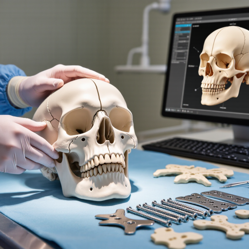
Assess the Fracture Pattern and Define Treatment Goals
How to Choose Cranio Maxillofacial Bone Plates?
Assess the Fracture Pattern and Define Treatment Goals
Start with the fracture map, not the plate catalog. Review imaging for fracture location, displacement, comminution, and nearby teeth, nerves, or sinus spaces. A clean fracture line differs from several small fragments. Note whether the bite has shifted and whether facial width or projection has changed. These details help the surgical team define what must be restored and what structures need protection.
Tips: Compare scans with the clinical exam. Record bite changes, tenderness, and visible asymmetry. Ask whether the planned fixation must resist substantial forces or mainly hold alignment. Small details matter.
Treatment goals should be specific: restore occlusion, facial contour, and stable bone contact while limiting unnecessary tissue disruption. Plate shape, thickness, and screw placement depend on the site, bone quality, and loading expected. Children and patients with weakened bone may need added consideration. A scan can look straightforward, yet the operative findings may differ. That uncertainty deserves a plan, not a guess. The treating surgeon should weigh imaging, examination, and experience before choosing fixation.
How to Choose Craniomaxillofacial Bone Plates
Assess the fracture pattern and define treatment goals
Illustrative relative planning-priority ratings (1 = lower emphasis, 5 = higher emphasis), not clinical outcome data or a treatment protocol. Plate choice depends on fracture location and complexity, bone quality, soft-tissue condition, and the clinician’s treatment goals.
Evaluate Anatomy, Bone Quality, and Mechanical Requirements
How to Choose Cranio Maxillofacial Bone Plates?
Evaluate Anatomy, Bone Quality, and Mechanical Requirements
Plate choice begins with the bone in front of you, not a catalogue. The orbital rim, zygomatic buttress, and mandible differ in thickness, curvature, and loading. Review CT images in multiple planes, then check whether the planned plate can sit flush without crowding a nerve, tooth root, or joint. Small details matter. A plate that bridges a gap may need to resist more bending than one supporting a well-aligned fracture.
Bone quality changes the fixation plan. The International Osteoporosis Foundation reports that one in three women and one in five men over 50 will experience an osteoporotic fracture. That statistic is not specific to facial bones, but it is a reminder to assess local bone purchase rather than assume uniform strength. The World Health Organization’s 2023 Global Status Report on Road Safety estimates 1.19 million road-traffic deaths annually, underscoring the varied forces behind facial trauma. Match screw length and plate rigidity to the site, fracture pattern, and expected load. Not a shortcut. A rigid construct is not automatically better; excessive stiffness or poor contour can complicate fit. Recheck the plan against imaging and the patient’s anatomy. Judgment still matters, and it is easy to underestimate that.
How to Choose Cranio Maxillofacial Bone Plates? — Evaluate Anatomy, Bone Quality, and Mechanical Requirements
| Anatomical area / use | Anatomy and mechanical demands | Bone-quality considerations | Typical plate and screw characteristics | Selection checks |
|---|---|---|---|---|
| Cranial vault fixation | Generally low-load fixation, but the plate must accommodate the curvature of the skull and avoid prominent hardware beneath thin soft tissue. | Assess local bone thickness and integrity, especially around fracture edges, prior surgical sites, and areas of thinning. | Low-profile plates and short screws are commonly used. Screw length must be appropriate for the local skull thickness and the specific system. | Confirm safe screw trajectory and length; avoid intracranial penetration. Check plate contour, edge prominence, and soft-tissue coverage. |
| Orbital rim and midface | Limited functional loading compared with the mandible. Thin bone, complex contours, and proximity to the orbit, teeth, and nerves constrain placement. | Bone may be thin or fragmented, with limited purchase at some fixation points. Consider the amount of intact bone available around the fracture. | Low-profile craniofacial plate systems are commonly selected; small-fragment systems often use screws around 1.2–2.0 mm in diameter, depending on the system and site. | Plan screw position and length from imaging and direct anatomy. Avoid the orbit, tooth roots, infraorbital canal, and other critical structures. |
| Zygomaticomaxillary complex | Multiple buttresses and fracture locations may require fixation at more than one point; plate contour and access vary by location. | Purchase can differ between the zygoma, maxillary buttress, and orbital rim. Comminution may reduce stable fixation options. | Low-profile plates are commonly used at selected fixation points. Plate size, hole pattern, and screw diameter depend on the anatomy and system. | Restore the intended three-dimensional alignment and check facial symmetry, orbital position, and plate fit before final fixation. |
| Mandible: symphysis and body | Subject to substantial forces from chewing and muscle activity. Fixation requirements depend on fracture pattern, location, occlusion, and whether load sharing is possible. | Assess cortical bone, fracture comminution, dental status, and the quality of bone available for screw purchase. | Small-fragment systems commonly include screw diameters around 2.0 mm; larger systems may use larger screws. Plate thickness and configuration vary with the required stability. | Consider occlusion, fracture stability, tooth roots, and the mandibular canal. Select fixation strategy according to the fracture and applicable clinical protocols. |
| Mandible: angle and ramus | Muscle forces and the fracture pattern can create rotational or bending forces. Access and the available bone corridor may limit plate placement. | Comminution, thin or compromised bone, and limited bone stock can reduce screw purchase and affect construct stability. | Plate configuration and screw size are system- and fracture-dependent. A low-profile option may suit some patterns; more robust fixation may be needed in others. | Evaluate fracture displacement, occlusion, canal location, and the planned screw trajectory. Do not assume the same construct is appropriate for every angle fracture. |
| Mandibular reconstruction or load-bearing fixation | May need to bridge a defect or highly unstable segment and carry greater functional loads than routine midface fixation. | Consider defect length, remaining bone stock, graft or flap plans, and whether the fixation points provide adequate purchase. | Typically requires a system designed for stronger load-bearing applications rather than a thin-profile midface plate. Plate dimensions and screw specifications vary substantially by system. | Plan construct strength, contour, and fixation points with the reconstructive team. Follow the device instructions and patient-specific surgical plan. |
| Pediatric craniofacial fixation | Growing anatomy and smaller structures require careful placement and consideration of future growth and development. | Bone dimensions and thickness differ from adults and vary with age and site. | Use fixation options specifically suitable for the patient’s age and anatomy; dimensions must be selected from the relevant system documentation. | Consider growth, tooth development, and nearby structures. Use age-appropriate imaging and specialist planning. |
| Poor-quality, osteoporotic, or compromised bone | Reduced purchase may increase the risk of loosening or loss of fixation, particularly where the construct is exposed to functional load. | Review imaging, bone stock, prior surgery, radiation history, infection, and other factors that may affect healing or fixation. | Plate choice alone may not compensate for inadequate bone purchase. Construct design and fixation strategy should be individualized. | Identify alternative fixation points or strategies as clinically appropriate; assess healing risks and follow-up needs. |
Important: Dimensions and screw sizes are system- and site-dependent; the ranges shown are illustrative, not a device prescription. Final selection and placement should be made by a qualified craniofacial surgeon using patient-specific anatomy, imaging, clinical judgment, and the applicable device instructions for use.
Compare Plate Materials, Shapes, and Fixation Options
Choosing cranio-maxillofacial plates means balancing strength, profile, and the bone’s healing needs. Titanium offers dependable rigidity and can be contoured for many fractures; resorbable polymer plates avoid a permanent metal implant but may have lower rigidity and require careful selection. Neither material suits every patient. Fit matters.
Shape should follow anatomy, not habit. Straight plates can stabilize simple lines, while L-, Y-, and orbital-specific plates help address angled or curved regions. Low-profile designs may reduce soft-tissue irritation, especially beneath thin facial tissue. ASTM F382-17 evaluates metallic bone-plate bending strength and stiffness using a four-point bend test. Those measurements help compare mechanical properties; they do not prove better clinical outcomes. A plate can pass a bench test and still be wrong for a complex fracture.
Fixation options matter just as much. Screw diameter, length, and number should match bone thickness and the planned construct; avoid placing screws where they may enter a tooth root or critical structure. Monocortical fixation may suit thinner bone, while bicortical purchase can provide stronger hold where anatomy permits. Not always. A neat plate diagram can hide awkward access or poor contouring. Surgeons should weigh imaging, fracture pattern, soft-tissue coverage, and healing risks, then document why a specific material and shape were chosen. References: ASTM F382-17; AO CMF Surgery Reference, fixation principles.
Match Plate Design to the Surgical Site and Technique
How to Choose Cranio Maxillofacial Bone Plates?
Match Plate Design to the Surgical Site and Technique
Plate choice starts with the anatomy, not the tray. A thin, curved plate may suit the orbital rim, where contour and limited space matter. The mandibular angle faces stronger chewing forces and often needs a more rigid construct. The Global Burden of Disease Study 2017 estimated 27.7 million new facial-fracture cases worldwide, underscoring how varied these injuries can be (GBD 2017 analysis of facial fractures). That figure describes fractures, not plate demand. Still, it highlights why one design cannot fit every site. Consider bone thickness, fracture pattern, access, soft-tissue coverage, and whether the planned approach allows accurate plate placement. A technically suitable plate can be awkward through a narrow incision. That detail is easy to underestimate.
Tips: Match plate shape and profile to the bone surface and surgical access. Check screw length against local bone thickness, and avoid placing hardware where it may interfere with tooth roots or delicate orbital structures. Follow current specialist guidance and the device’s instructions for use. A tidy plan may still need revision in the operating room; anatomy does not always match the image.
Confirm Device Compatibility, Safety, and Clinical Evidence
Choosing cranio-maxillofacial bone plates starts with confirming that the plate, screws, instruments, and planned anatomy work together. Review CT images for bone thickness, fracture lines, and nearby structures such as the orbit, teeth, and sinuses. Check screw diameter and length against the intended fixation points; a plate that needs forceful bending may not sit as planned. Fit matters. Confirm the material and its documentation, too. ASTM F136 specifies titanium-6aluminum-4vanadium ELI alloy for surgical implant applications, but the alloy name alone does not establish suitability for every case. Follow the device’s validated instructions for use, including sterilization and imaging considerations.
Safety claims should be supported by test documentation and relevant clinical evidence, not a product description alone. The CDC reports that surgical site infections occur in about 2–4% of patients undergoing inpatient surgery; this is a broad figure, not a craniofacial plate-specific rate. Compare studies that report infection, loosening, exposure, and reoperation, and check whether their patients and follow-up periods resemble the intended use. Evidence can be uneven. Small case series may miss late problems, while pooled results can combine different injuries and techniques. One useful question is whether the evidence measures outcomes that matter in practice, such as stable fixation and uncomplicated healing, rather than simply counting implanted devices. Local review of imaging, operative records, and follow-up can reveal gaps that published averages cannot.
FAQS
Review imaging for fracture location, displacement, and small bone fragments. Check nearby teeth, nerves, and sinus spaces.
A clean fracture line differs from several loose fragments. The pattern helps determine how much support is needed.
The team may aim to restore the bite, facial contour, and stable bone contact. Less tissue disruption is also a goal.
The orbital rim, cheekbone support, and jaw differ in thickness and curvature. A plate should sit flush without crowding nearby structures.
Yes. Weaker bone may provide less secure screw purchase. Age and health can matter, but each patient is different.
No. Rigidity must match the fracture and expected forces. Excessive stiffness or poor fit can create problems.
Consider the fracture site, bone quality, and nearby tooth roots or nerves. Screw length should suit the available bone.
Not always. Operative findings may differ, so the plan may need adjustment. That uncertainty is real.
Conclusion
Choosing Cranio Maxillofacial Bone Plates begins with understanding the fracture pattern and defining clear treatment goals, such as restoring alignment, stability, and function. The surgeon should assess the affected anatomy, bone quality, and forces the fixation will need to withstand. These factors help determine whether a particular plate’s material, shape, thickness, and fixation options are appropriate for the case.
The plate design should also suit the surgical site and planned technique, allowing practical placement while supporting the intended repair. Before selection, confirm that the plate and screws are compatible with one another and with any relevant instruments, and review applicable safety information and clinical evidence. Considering these points together supports a careful, case-specific choice rather than relying on a single feature.

Blog Tags: