Auxein Medical Orthopedic & Clinical Blog Banner
Insights & Innovation

Orthopedic Insights & Knowledge

Explore the latest advances in orthopedic implants, trauma solutions, surgical technology, and healthcare industry news from Auxein Medical.

0%

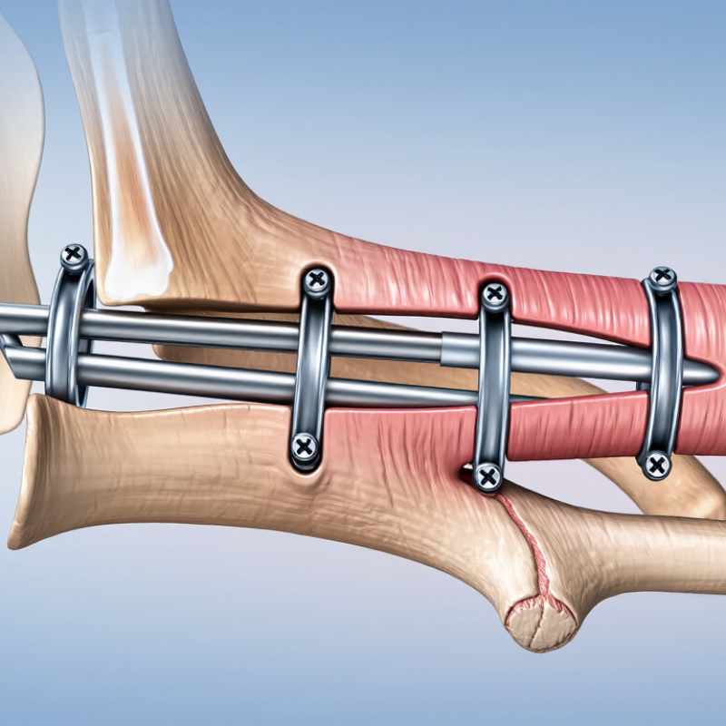
A Tibia Intramedullary Nail is a metal implant placed inside the tibia’s central canal to stabilize certain fractures. Surgeons typically insert it through an incision near the knee, guide it along the bone, and secure it with locking screws. On an X-ray, the nail appears as a slender rod running through the shinbone, with screws crossing it near the fracture.

The choice of fixation depends on the fracture’s location, pattern, and stability, as well as the patient’s overall health. Orthopedic trauma specialist Peter V. Giannoudis has published extensively on fracture care, but I cannot verify a verbatim quotation from him about tibial nails. A fabricated quote would be misleading. His work supports a careful, evidence-based approach: the implant must suit the injury, not the other way around.

This guide explains how a Tibia Intramedullary Nail works, when clinicians may consider it, and what recovery can involve. It also covers possible complications, including infection, delayed healing, and irritation around the knee. Details matter. A nail can provide strong internal support, but it does not make every fracture simple, and healing timelines vary. Patients should discuss personal treatment decisions with their orthopedic team.
What Is a Tibia Intramedullary Nail?

Definition and Purpose of a Tibial Intramedullary Nail

What Is a Tibia Intramedullary Nail?

A tibial intramedullary nail is a metal rod placed inside the hollow center of the tibia, or shinbone. Surgeons use it to stabilize certain fractures, especially breaks along the bone’s shaft. The nail follows the bone’s natural channel, while locking screws near its ends help limit unwanted movement. It acts as internal support while healing progresses; it does not instantly repair the fracture. The exact plan depends on the break’s location, pattern, and the patient’s health.

The purpose is to keep broken bone sections aligned and steady enough for healing. In suitable cases, this fixation can help patients begin guided movement earlier than some other methods. But recovery varies. Pain, swelling, and limited weight-bearing may still occur, and follow-up imaging helps clinicians assess healing. A nail is not the right choice for every tibial fracture. That distinction matters.

Tips: Ask your orthopedic clinician where the fracture is, how the nail will be secured, and what weight-bearing limits apply. Keep follow-up appointments, even when the leg feels better. Healing can be less obvious than expected. An individual treatment plan should come from a qualified medical professional.

Tibial Anatomy and Fractures Treated With Intramedullary Nailing

A tibial intramedullary nail is a metal rod placed inside the tibia’s central canal to stabilize selected fractures. The tibia is the larger, weight-bearing bone of the lower leg. Its shaft has a relatively limited soft-tissue covering, especially along the front edge, so fractures can involve skin and surrounding tissue as well as bone. The nearby fibula, knee, and ankle also matter when assessing alignment and injury.

The fracture pattern guides treatment. Intramedullary nailing is commonly used for many tibial shaft fractures, including some caused by high-energy impacts. The surgeon passes the nail through the canal and secures it with screws above and below the break. Those screws help control rotation and length. A 2006 epidemiological review by Court-Brown and Caesar reported an estimated tibial shaft fracture incidence of 16.9 per 100,000 people annually. That figure describes population burden, not an individual patient’s risk. Anatomy is rarely tidy: fractures may extend toward the knee or ankle, and open wounds can complicate care. Nail placement also requires attention to alignment, because a small angular error can affect how the leg bears weight. Intramedullary nailing is not right for every tibial fracture; clinical examination and imaging help determine whether it fits.

Design, Materials, and Main Components of the Implant

What Is a Tibia Intramedullary Nail?

A tibial intramedullary nail is a metal implant placed inside the hollow center of the shinbone. It supports alignment while the fracture heals. Unlike an external frame, the nail sits beneath the skin and shares load with the bone. Its shape must match the tibia’s narrow canal, gentle curve, and changing diameter.

Most nails use titanium alloy or stainless steel. Titanium is light, strong, and relatively flexible, while stainless steel offers high strength and predictable imaging characteristics. The main components include the nail body, proximal locking holes, distal locking holes, and an end cap. Locking screws pass through the bone and nail. They control rotation, shortening, and movement. Some designs include multiple screw directions near the knee or ankle. This improves control in complex fractures, but it may also make surgery more demanding.

Tips: Nail length and diameter should match the patient’s anatomy and fracture pattern. Careful imaging helps confirm the entry point, canal size, and screw position. Surgeons should also check soft tissues and nearby joints. No design is perfect. A nail that works well in one tibia may not suit another. Small anatomical differences matter, and clinical judgment remains essential. Poor alignment can affect walking, even when the implant appears secure.

Tibia Intramedullary Nail: Materials and Structural Design

Tibial intramedullary nails are load-sharing implants placed inside the medullary canal. The nail body is commonly manufactured from titanium alloy or stainless steel, while locking screws secure alignment and control rotation and length. The chart compares representative minimum yield-strength values for commonly used implant metals.

Representative engineering values: titanium alloy Ti-6Al-4V ≈ 830 MPa, 316L stainless steel ≈ 170 MPa, and cobalt-chromium-molybdenum alloy ≈ 450 MPa. Actual values vary with composition, heat treatment, manufacturing process, and implant design.

Main implant components:
  • Nail body: a hollow or solid longitudinal implant that supports alignment and shares load with the tibia.
  • Locking screws: transverse screws that limit rotation and telescoping.
  • End cap: closes the proximal or distal opening and may help control nail position.
  • Design features: insertion geometry, locking holes, curvature, diameter, and length are selected according to anatomy and fracture pattern.

How Tibial Intramedullary Nail Surgery Is Performed

What Is a Tibia Intramedullary Nail?

How Tibial Intramedullary Nail Surgery Is Performed

A tibial intramedullary nail is a metal rod placed inside the shinbone. It stabilizes a fractured tibial shaft while new bone develops. Epidemiological research reports tibial shaft fractures at roughly 17 cases per 100,000 people each year. Treatment depends on fracture location, skin damage, alignment, and the patient’s general health.

During surgery, the patient receives regional or general anesthesia. The surgeon makes a small incision near the knee or, sometimes, near the ankle. Fluoroscopy provides live X-ray images. A guidewire passes through the marrow canal and crosses the fracture. The canal may be gently widened. The nail then follows the wire into the tibia. Locking screws secure both ends and control rotation. A real detail matters here: the final alignment is checked from several angles, not just one.

The American Academy of Orthopaedic Surgeons’ clinical guidance supports intramedullary nailing for many displaced tibial shaft fractures. NICE guidance also recognizes it as a standard surgical option when appropriate. Surgery often permits earlier weight bearing than prolonged casting, but recovery is not perfectly predictable. Infection, knee pain, compartment syndrome, delayed union, and malalignment can still occur. A Cochrane review has described the evidence for some technique choices as limited or low certainty. That uncertainty deserves honest discussion.

What Is a Tibia Intramedullary Nail? — How Tibial Intramedullary Nail Surgery Is Performed
Data Dimension Key Information Clinical Details Typical Considerations
Definition A tibial intramedullary nail is a metal rod placed inside the hollow central canal of the tibia. The implant helps align and stabilize a broken shinbone while the fracture heals. It is commonly secured with locking screws placed through the bone and nail.
Main Fracture Location Most often used for fractures of the tibial shaft, the long middle portion of the shinbone. It may also be considered for selected fractures near the upper or lower ends of the tibia, depending on the fracture pattern. Fracture location, alignment, soft-tissue condition, and associated injuries influence treatment selection.
Common Indications
  • Displaced or unstable tibial shaft fractures
  • Comminuted fractures with multiple bone fragments
  • Some open fractures after appropriate wound management
  • Fractures that are poorly suited to casting or bracing
The procedure is selected when internal stabilization is expected to improve alignment, mobility, or fracture healing. Not every tibial fracture requires an intramedullary nail; treatment is individualized after imaging and examination.
Preoperative Assessment Evaluation usually includes a physical examination and imaging studies. X-rays are standard; computed tomography may be used for complex fractures or fractures involving a joint. Clinicians assess circulation, sensation, swelling, skin condition, infection risk, and other injuries.
Anesthesia Surgery is performed with anesthesia to control pain and prevent movement. General anesthesia or regional anesthesia may be used, depending on the patient, operation, and anesthesiology plan. Medication history, medical conditions, allergies, and fasting instructions are reviewed before surgery.
Patient Positioning The patient is positioned to allow access to the knee and lower leg. The leg is prepared and draped using sterile technique. Fluoroscopy is commonly used to view the fracture and implant during the operation. The surgical approach may vary according to the fracture location and the surgeon’s preferred technique.
Entry Point A small incision is made near the upper end of the tibia, commonly through an approach near the knee. An entry guide is placed into the tibial canal under imaging guidance. Care is taken to protect the knee structures and to establish a correct path through the medullary canal.
Canal Preparation The medullary canal may be opened and gradually prepared to accommodate the nail. In reamed techniques, flexible instruments enlarge the canal in measured steps. Some fractures may be treated without reaming. The chosen method depends on fracture characteristics, bone anatomy, soft-tissue status, and surgical judgment.
Fracture Reduction The bone fragments are realigned before or during nail insertion. Traction, positioning, temporary instruments, or small additional incisions may be used to restore length, rotation, and alignment. Accurate reduction is important because malalignment can affect walking mechanics and lower-limb function.
Nail Insertion The nail is advanced through the tibial canal across the fracture site. Imaging is used to confirm the nail’s position, fracture alignment, and appropriate depth. The nail is selected to match the patient’s tibial anatomy and the fracture requirements.
Locking Screws Screws are inserted through holes in the nail to control rotation and maintain length. Locking may be performed at both ends of the nail. The number and direction of screws depend on the fracture pattern and nail design. Stable fixation helps maintain alignment during early healing and rehabilitation.
Wound Closure Incisions are irrigated and closed after final imaging confirms satisfactory fixation. Dressings are applied, and the limb is monitored for swelling, bleeding, circulation, and sensation. The operation may be followed by observation in a recovery area before discharge or hospital admission.
Weight Bearing Weight-bearing instructions vary according to fracture stability, bone quality, soft-tissue condition, and fixation. Some patients begin partial or progressive weight bearing early, while others require restricted weight bearing. Patients should follow the surgeon’s specific instructions and use crutches or a walker when prescribed.
Rehabilitation Rehabilitation commonly focuses on knee and ankle motion, swelling control, strength, balance, and walking. Physical therapy may be recommended to restore movement and muscle function. Progression is guided by symptoms, examination, radiographs, and evidence of fracture healing.
Healing and Follow-Up Follow-up visits monitor the incision, implant position, alignment, and development of bridging bone. Healing time varies substantially with fracture type, soft-tissue injury, smoking status, medical conditions, and treatment factors. Radiographic healing does not always occur at a fixed time; activity is advanced according to clinical and imaging findings.
Potential Benefits Internal fixation can provide strong stabilization while preserving alignment along much of the tibial shaft. Compared with prolonged casting in suitable cases, it may facilitate earlier movement of the knee and ankle and support functional recovery. Expected benefits must be balanced against surgical risks and the patient’s overall health.
Possible Complications
  • Infection
  • Bleeding or blood clots
  • Delayed union or nonunion
  • Malalignment or rotational deformity
  • Knee or ankle stiffness
  • Anterior knee pain
  • Implant irritation, breakage, or need for later removal
  • Nerve or blood-vessel injury
Risk varies with the injury, open wounds, smoking, diabetes, bone quality, and other health factors. Increasing pain, fever, drainage, calf swelling, numbness, color change, or shortness of breath requires prompt medical attention.
When Implant Removal May Be Considered Removal is not routinely required after healing. It may be considered for persistent implant-related pain, prominent hardware, infection, or other specific clinical reasons. Removal is another surgical procedure and is recommended only when potential benefits outweigh its risks.

Recovery, Benefits, Risks, and Possible Complications

What Is a Tibia Intramedullary Nail?

A tibial intramedullary nail is a metal rod placed inside the shinbone’s hollow center. Surgeons use it to stabilize many unstable or displaced tibia fractures. Locking screws hold the bone and nail in position while healing progresses. This method often supports earlier movement than prolonged casting, but weight-bearing depends on fracture stability and surgical findings.

Recovery usually begins with swelling control, wound checks, and gentle ankle and knee exercises. A physical therapist may help you move from a walker to crutches, then toward normal walking. Some patients bear weight early. Others need several weeks of limited loading. Healing commonly takes months, and follow-up X-rays guide each change. Progress is rarely perfectly linear. It can be humbling.

The procedure offers strong alignment and protects the fracture during daily movement. However, risks include infection, bleeding, blood clots, nerve irritation, and damage to nearby tissues. Persistent knee pain near the entry point is also reported by some patients. Delayed healing, nonunion, malalignment, or broken locking screws may require further treatment. Increasing redness, drainage, fever, calf swelling, numbness, or worsening pain needs prompt medical attention. Your orthopedic team should adjust activity levels using examination, imaging, and your actual symptoms. Pain alone cannot always show whether the bone has healed.

FAQS

What is a tibial intramedullary nail?

It is a metal rod placed inside the shinbone’s hollow center. It helps stabilize a fracture while bone heals.

What materials are commonly used?

Nails are usually made from titanium alloy or stainless steel. Titanium is lighter and relatively flexible; stainless steel offers high strength.

What parts make up the implant system?

The system includes a nail body, locking holes, screws, and an end cap. Screws pass through the bone and nail to limit rotation and shortening.

How does the surgeon place the nail?

A guidewire is passed through the tibial canal and across the fracture. The nail follows the wire, then screws secure both ends.

How does the surgical team check alignment?

Live X-ray images help guide placement. The final alignment is checked from several angles, since one view may miss a problem.

What affects the choice of nail size?

The tibia’s length, canal width, curve, and fracture pattern all matter. A nail that fits one person may not suit another.

Can surgery allow earlier weight bearing than casting?

It often can, but recovery varies. The patient’s fracture, overall health, and healing progress influence when weight bearing is safe.

What complications may occur?

Possible problems include infection, knee pain, compartment syndrome, delayed healing, and poor alignment. A secure-looking implant does not guarantee perfect walking.

Is the best surgical technique settled?

Not entirely. Some evidence about technique choices remains limited or uncertain, so patients and surgeons should discuss trade-offs openly.

Conclusion

A Tibia Intramedullary Nail is a metal implant placed inside the central canal of the shinbone to stabilize a fracture while the bone heals. It is commonly used for breaks in the tibial shaft and certain fractures near the ends of the bone. The tibia bears much of the body’s weight, so restoring its alignment and stability is important for safe movement and recovery. The nail is typically secured with screws that help prevent unwanted rotation or shortening.

Surgery generally involves inserting the nail through a small opening near the knee or ankle, positioning it within the bone, and fixing it in place with screws. Implants are made from strong, biocompatible materials and come in designs suited to different fracture patterns and patient needs. Compared with some alternatives, this method can provide stable fixation and support earlier movement, though recovery varies. Possible risks include infection, blood clots, irritation around the entry point, delayed healing, or the need for further treatment. Follow-up care and guided rehabilitation help monitor healing and restore strength and mobility.

Evelyn

Evelyn

Evelyn is a dedicated marketing professional with a strong understanding of the company’s products, services, and customer-focused solutions. Through clear communication and thoughtful content strategy, she helps translate complex product features into practical insights that are easy for readers......