Orthopedic Insights & Knowledge
Explore the latest advances in orthopedic implants, trauma solutions, surgical technology, and healthcare industry news from Auxein Medical.
Why Choose Cannulated Bone Screws?
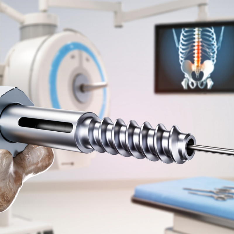
Cannulated Bone Screws are designed with a hollow central channel. This channel allows surgeons to place the screw over a guidewire. The technique can improve accuracy during fracture fixation, especially when access is limited or the target area is deep. In the operating room, the guidewire acts like a narrow path through bone. Fluoroscopy helps confirm its position before drilling and insertion.
Orthopedic trauma surgeon Dr. Paul Tornetta III has expressed a practical principle: “The guide wire establishes the trajectory; the screw follows it.” This idea explains the main appeal of Cannulated Bone Screws. They support controlled placement while reducing repeated attempts at alignment. That matters near joints, where a few millimeters can affect stability and movement. Still, the system is not automatically safer. A guidewire can bend, migrate, or sit in an incorrect position. Careful imaging and surgical judgment remain essential.
Material choice, thread design, diameter, and compression also influence performance. A partially threaded screw may create interfragmentary compression, while a fully threaded option can support different fixation strategies. Patient bone quality matters too. Osteoporotic bone may require another construct or additional fixation. This is where simplified product claims become unreliable. The best choice depends on fracture pattern, anatomy, surgeon experience, and imaging control. Cannulated Bone Screws offer precision, but precision requires discipline. Their value comes from proper planning, not from the hollow design alone.
What Are Cannulated Bone Screws?
Why Choose Cannulated Bone Screws?
What Are Cannulated Bone Screws?
Cannulated bone screws are hollow orthopedic implants. Their central channel accepts a thin guidewire during surgery. The surgeon places the wire first, checks its position with imaging, then advances the screw along the planned path. This method can improve trajectory control, especially around the hip, ankle, wrist, and small bone fragments.
The concept is elegant, but not flawless. A hollow core may reduce some mechanical strength compared with a solid screw of similar size. Guidewire migration, bending, or breakage can also create avoidable problems. Careful imaging and controlled drilling remain essential. The instrument does not replace judgment.
The Global Burden of Disease 2019 study estimated 178 million new fractures worldwide in one year. It also reported about 455 million people living with fracture-related health consequences (Lancet Healthy Longevity, 2021). These figures explain the demand for precise fixation, but they do not prove that cannulated screws suit every fracture. Comparative orthopedic reviews generally show that outcomes depend on fracture pattern, bone quality, screw placement, and surgeon experience. I would not call the design automatically superior. Its real value is practical: guided insertion, smaller access points, and more predictable alignment when the indication is appropriate.
Why Choose Cannulated Bone Screws? - What Are Cannulated Bone Screws?
| Data Dimension | Typical Information | Clinical Relevance |
|---|---|---|
| Definition | A cannulated bone screw is a fixation screw manufactured with a hollow central channel extending through part or all of its length. | The channel allows the screw to be guided over a thin guidewire or guide pin during placement. |
| Guided Insertion | The guidewire is positioned first, and the screw is advanced over it to the intended location. | This technique can help the surgeon control the entry point, trajectory, and final screw position, particularly in minimally invasive procedures. |
| Common Materials | Common implant materials include titanium alloys and stainless steel alloys intended for surgical implants. | Material selection affects strength, corrosion resistance, imaging characteristics, stiffness, and compatibility with the surgical system. |
| Common Screw Diameters | Frequently used systems include approximate outer diameters of 2.0 mm, 2.4 mm, 3.0 mm, 3.5 mm, 4.0 mm, 4.5 mm, 6.5 mm, and 7.3 mm. Available sizes vary by indication and system. | A wider range of diameters allows selection according to bone size, fracture pattern, required purchase, and soft-tissue considerations. |
| Available Lengths | Typical systems offer multiple lengths, often in increments of approximately 1–5 mm, depending on the diameter and design. | Multiple lengths help achieve appropriate bicortical or subchondral fixation while reducing the risk of excessive screw prominence. |
| Thread Design | Designs may include fully threaded, partially threaded, or variable-pitch threads. Thread profiles can be coarse or fine according to the intended bone and fixation method. | Thread configuration influences compression, pull-out resistance, fragment control, and the ability to achieve lag fixation. |
| Head Configuration | Heads may be countersunk, partially threaded, low-profile, or designed to accept a washer. Recesses commonly support a dedicated driver. | Head geometry affects soft-tissue irritation, load distribution, driver engagement, and the ability to seat the screw flush with the bone surface. |
| Guidewire Compatibility | Each screw system is designed for a specified guidewire diameter and insertion instrument. The guidewire must match the system requirements. | Correct compatibility supports accurate placement and helps prevent guidewire-screw mismatch or obstruction during insertion. |
| Compression Capability | Partially threaded screws can provide interfragmentary compression when the threads engage the far fragment while the near fragment is allowed to glide. | Compression can improve fracture stability and promote contact between appropriately prepared bone surfaces. |
| Typical Applications | Applications may include fracture fixation, arthrodesis, osteotomy fixation, and fixation of small or large bone fragments, depending on the screw design. | The same guided-placement principle can be adapted to different anatomical regions when the implant size and fixation strategy are appropriate. |
| Common Anatomical Uses | Examples include selected fractures of the foot, ankle, wrist, hand, hip, and other skeletal regions where accurate screw trajectory is important. | Anatomical use depends on bone quality, fragment size, fracture geometry, implant dimensions, and the treating surgeon’s plan. |
| Potential Advantages | Key advantages include guidewire-based placement, improved trajectory control, access through smaller approaches, and availability in multiple thread and size configurations. | These characteristics may support accurate fixation while limiting unnecessary dissection, although outcomes depend on patient factors and surgical technique. |
| Potential Limitations | The hollow channel can reduce the solid cross-sectional area of the screw. Guidewire bending, migration, breakage, or retained wire fragments are also procedural concerns. | Careful handling, appropriate instrumentation, correct sizing, and adherence to the implant’s surgical technique are important for safe use. |
| Bone Quality Considerations | Poor bone quality may reduce thread purchase and pull-out resistance, regardless of whether the screw is cannulated. | The fixation plan may require a different screw size, a washer, supplemental fixation, or another construct based on the clinical situation. |
| Imaging and Verification | Fluoroscopy or other intraoperative imaging is commonly used to confirm the guidewire path, screw length, joint clearance, and final implant position. | Image verification is especially important when working near articular surfaces, neurovascular structures, or narrow anatomical corridors. |
| Selection Checklist | Selection should consider diameter, length, thread pattern, head design, material, guidewire size, bone quality, fracture pattern, and intended compression. | Choosing the correct configuration helps balance stability, tissue protection, insertion control, and the risk of implant prominence or inadequate fixation. |
FAQS
They contain a hollow channel through the center. This channel allows placement over a guidewire.
The guidewire helps maintain the planned entry point and trajectory. Surgeons can check alignment before final insertion.
They may help with small fragments, narrow spaces, and anatomically complex areas. Precision matters.
Suitable designs can create compression across a fracture. This may improve contact between bone surfaces.
No. Their benefits depend on fracture pattern, bone quality, screw design, and surgical technique.They are not magic.
Diameter should match bone size and mechanical demands. Length should cross the fracture without unnecessary protrusion.
Poor bone quality may require another fixation strategy or additional support. Aggressive compression may not be appropriate.
Imaging helps confirm guidewire direction and final depth. More than one view can reveal misleading alignment.
A bent guidewire, poor imaging angle, or incorrect screw length can reduce fixation quality. Small errors matter.
They should assess soft tissues, healing potential, patient-specific risks, and the surgical approach. Careful training remains essential.The simple plan may fail.
Conclusion
Cannulated Bone Screws are specialized orthopedic implants designed with a hollow central channel that allows surgeons to guide the screw over a temporary wire during placement. This design supports accurate alignment, controlled insertion, and reliable fixation of bone fragments. By combining a guided technique with strong compression, these screws can help stabilize fractures and support proper healing while reducing unnecessary disruption to surrounding tissue.
They are commonly used in procedures involving fractures of the hip, ankle, wrist, foot, and other areas where precise fixation is important. Key benefits may include improved placement accuracy, efficient surgical handling, and effective compression across the fracture site. Selecting the right Cannulated Bone Screws requires consideration of bone quality, fracture pattern, screw diameter and length, thread design, required compression, and the patient’s overall clinical needs. These factors help surgeons choose a fixation solution suited to each procedure.